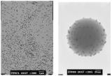
| Reporting in Journal of Nanomaterials, Bergman and colleagues described a method to make fluorescent dye functionalized silica nanoparticles with a series of surface functionalization steps, and established the relationship between particle suspension stability with surface functional group of the particles that were prepared. |
Reviewed by Li Han, RTI International
- Bergman, et al. (2008) " On the Complexity of Electrostatic Suspension Stabilization of Functionalized Silica Nanoparticles for Biotargeting and Imaging Applications," J. Nanomater. v2008 (2008) articleID 712514. DOI: 10.1155/2008/712514
Nanoparticles have attracted tremendous attention as fluorescent markers in biological applications since the size of these materials enables them to cross physiological barriers for efficient drug delivery and medical diagnosis. While the most commonly used nanoparticles for this purpose are polymer based, their size, surface function, and surface charge controllability remain a challenge. Inorganic materials such as silica particles are emerging as a promising alternative to polymer particles due to their highly tunable particle size and surface chemistry. Reporting in Journal of Nanomaterials, Bergman and colleagues describe a method to make fluorescent dye functionalized silica nanoparticles with a series of surface functionalization steps, and establish the relationship between particle suspension stability with surface functional group of the particles that were prepared.
In this report, the Streptavidin Alex Fluor 55 functionalized silica particles were prepared by three steps, including silica particle synthesis, surface functionization, and bioconjugation of fluorescent dye labeled protein on particle surface. In their approach, the silica particles were synthesized using Stöber synthesis method, followed by surface functionalization of polyethyleneimine (PEI) with aziridine to serve as precursor. The PEI silica particles then react with succinic anhydride or glutaraldehyde to form carboxylic acid group and aldehyde group that will serve as medium for subsequent streptavidin conjugation. Three types of nanoparticle systems are produced, PEI-SiO2; glutaraldehyde-PEI- SiO2 and succinic acid–PEI- SiO2. The properties of the silica particles are evaluated with dynamic light scattering and microscopy to determine the particle size and shape. The isoelectric point (IEP) of silica particle with different functional groups was obtained by electrokinetic titrations. Gold particles were used as labeling agent to allow easy identification of the streptavidin in TEM imaging.

With the bioconjugation of the Alexa Fluor 55-streptavidin on the particle surface, confocal fluorescence microscopy confirmed that the PEI- SiO2 suspension had the least agglomeration compared with two other systems, which is in good agreement with the experimental zeta potential–pH relation for the three particle systems, where PEI- SiO2 has the highest zeta potential at pH 6. The presence of the streptavidin on SiO2 particles was also confirmed by gold particle labeling in TEM imaging, where again a large degree of agglomeration for glutaraldehyde–PEI- SiO2 particles and succinic acid-PEI- SiO2 particles was observed.
The PEI-functionalized silica particles are found to be very promising candidates for biological application due to their positive charge carrying capability and better suspension formation in neutral pH. Furthermore, the understanding of the relationship between particle surface zeta potential and surface functionalization will have important implication on designing particles with high suspension stability for drug delivery and clinical diagnosis applications.
Image from Bergman, et al. (2008) "On the Complexity of Electrostatic Suspension Stabilization of Functionalized Silica Nanoparticles for Biotargeting and Imaging Applications", J. Nanomater. v2008 (2008) articleID 712514. Reused under a Creative Commons Attribution License.
This work is licensed under a Creative Commons Attribution-NonCommercial-NoDerivs 3.0 Unported.
